CrysAlisᴾʳᵒ: An All-in-one Software Package for Single Crystal X-ray Diffraction
Introduction
 CrysAlisPro is the dynamic data collection and data processing program supplied with Rigaku Oxford Diffraction systems. It is designed with an intuitive and user-friendly graphical user interface that runs in fully automatic, semi-automatic or manual modes. At the heart of the program are four modules that take the user through automatic crystal screening, strategy calculation, data collection, and data processing. CrysAlisPro emphasizes live feedback, such as crystal quality and data statistics throughout sample screening and data collection, and guides the user on the best exposure time and image rotation width. Furthermore, CrysAlisPro integrates control of system components, making it possible to run the entire crystallography experiment from one program.
An all-in-one interface:
Cryotemperature setup, X-ray generator control, beam divergence tuning, data collection and processing
CrysAlisPro gives users complete control of their macromolecular crystallography experiments, from setting up the X-ray generator and cryosystem to data collection and processing. To demonstrate these capabilities, we show how CrysAlisPro was used to perform all the steps of an X-ray analysis for a thaumatin crystal. For this study, a Rigaku Oxford Diffraction XtaLAB Synergy-S system was configured with a microfocus PhotonJet-S sealed-tube source, a universal 4-circle kappa goniometer and a HyPix6000-HE Hybrid Photon Counting detector.
Cryotemperature setup:
The CrysAlisPro software controls various cryosystems, including the Oxford Cryostream Series and the Cobra. In addition to setting up any desired temperature set-point, users can also opt to automatically warm up the cryosystem to room temperature after data collection. All programs pertaining to the cryosystem, such as purging the system for maintenance or varying the temperature in steps, are also available from CrysAlisPro as shown in Figure 1. For these experiments, the cryosystem temperature target of 100K was selected and the “Cool” program was run. After achieving the set temperature, the thaumatin crystal was mounted on the goniometer and then centered in the X-ray beam for data collection.
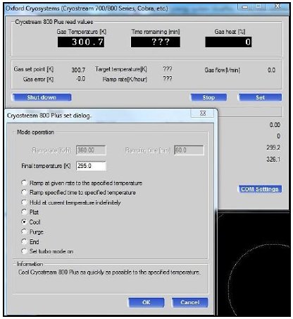
Figure 1: The cryosystem control interface.
Crystal Screening:
CrysAlisPro provides two options for crystal screening. The first option collects a “Single scan” of data consisting of consecutive images. The other collects “Two scans” of data consisting of scans that collect images 90° apart. The number of images per scan and the angle between scans are configurable (Figure 2).
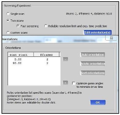
Figure 2: Screening strategies interface.
With one mouse click, CrysAlisPro automatically runs the following series of steps without any other user intervention:
- Ramp up the XtaLAB Synergy S X-ray source to full power. Users can also opt to have CrysAlisPro automatically ramp down the X-ray power if the system remains unused for a user-defined period of time. This greatly helps to prolong the lifetime of the X-ray tube.
- Collect and concurrently index the diffraction images and show resulting unit cell parameters, Bravais lattice and the number and percentage of indexed reflections.
- Analyze the crystal quality by estimating the resolution limit for which I/σ(I) equals 2 and the crystal mosaicity in the three spatial dimensions.
For these experiments, a thaumatin crystal was screened using the “Single Scan” option with an exposure time of 1 second per 0.3°. Figure 3 shows the results of indexing. After screening, the reciprocal lattice viewer was used to verify how well reflections intersected with crystal lattice points, as well as the intensity distribution along the a*, b* and c* axes (Figure 4).
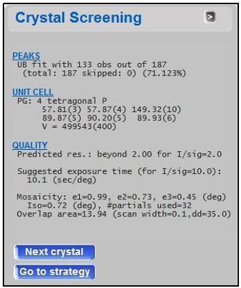
Figure 3: Screening results for the thaumatin crystal.
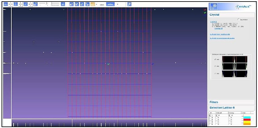
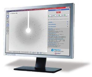
Figure 4: Reciprocal lattice viewer
Beam Divergence Tuning:
In cases where samples exhibit long unit cell parameters and overlapping reflections, it is critical to reduce the reflection size by reducing the beam divergence. CrysAlisPro seamlessly controls the beam divergence slit for XtaLAB Synergy and DW diffractometers. A simple interface (Figure 5) allows users to tune the beam divergence (between ~9.2 milliradians and 1 milliradian) as needed to separate reflections.
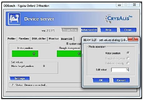
Figure 5: Divergence slit control.
For the thaumatin crystal in this study, a beam divergence of ~6.5 mrad was sufficient to resolve reflections along the 150 Å axis at a crystal to detector distance of 35 mm, as shown in Figure 6. By using the slits to tune divergence, one can get optimal resolution of reflections without sacrificing measurement resolution at the edge of the detector
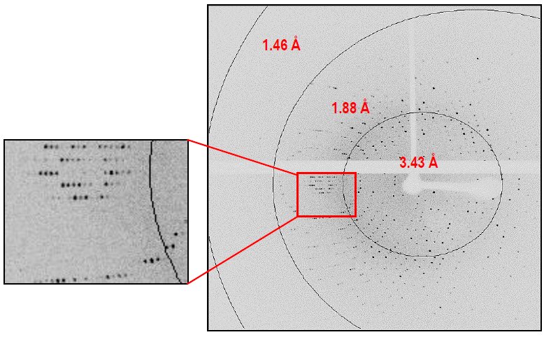
Figure 6: Representative diffraction image for thaumatin, collected at 35 mm with slit setting of ~6.5 mrad.
Strategy Calculation and Data Collection:
CrysAlisPro has a comprehensive strategy calculation module (Figure 7) with the following features to make the process simple and efficient:
- Users can readily set the resolution target, select the Laue group and choose whether to merge the Friedel mates.
- Several types of strategy are accessible through a pull-down menu. The default strategy calculates a complete data set, regardless of redundancy. Some other options are (a) to calculate a complete data set with user-selected target redundancy, (b) collect a data set (even if incomplete) within a user-selected time constraint, and (c) collect a data set within a user-selected time constraint and with an adjusted exposure time to reach a complete data set.
- For each new strategy, CrysAlisPro estimates the exposure time necessary to reach a user-specified I/σ(I) = 2 at the selected resolution target.
- CrysAlisPro determines the optimal rotation width to avoid overlaps due to crystal mosaicity and unit cell dimensions.
- CrysAlisPro displays estimated completeness and redundancy statistics in both the Laue group determined upon indexing and in P1.
- An advanced dialog offers additional options, including data collection along a selected symmetry axis or to collect along the longest unit cell axis first. In this dialog, users can also define a cryonozzle shadow on the diffraction images or implement constraints on the goniometer axes.
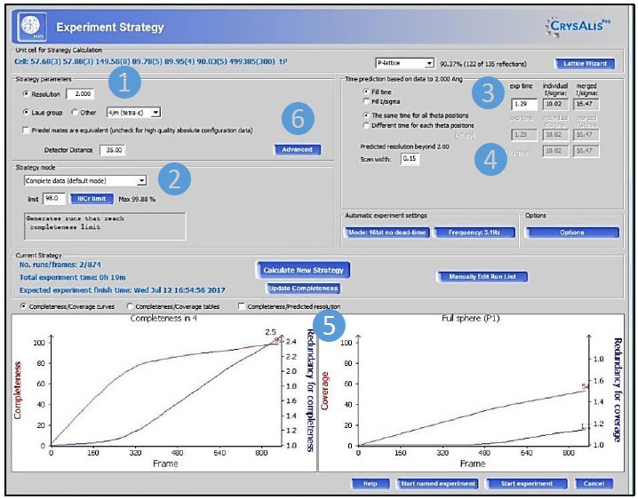
Figure 7: Strategy calculation interface
A strategy was calculated for this thaumatin crystal using CrysAlisPro and a crystal to detector distance of 35 millimeters. The default “Complete Data Set” option was selected to achieve data completeness to 1.50 Å with an overall <I/σ(I)> of 15 for the Friedel pairs unmerged. Two scans were calculated by CrysAlisPro with an exposure time of 4 sec per 0.15º (Table 1). The beam divergence was kept at ~6.5 mrad.
Table 1: Scans collected on thaumatin.
| Exp | Δω | Dist | 2θ | φ | κ | # img | Timeexp |
| 4 sec | 0.15º | 35 mm | -15.17° | -87º | 109º | 354 | 23m 36s |
| 4 sec | 0.15º | 35 mm | -15.17° | -171º | 40º | 314 | 20m 56s |
| 44m 32s | |||||||
The data were concurrently and automatically processed by CrysAlisPro during data collection to 1.50 Å using all default parameters. The results, in Table 2, show that data completeness to 1.50 Å was 98.6% and Rint = 6.8% . Furthermore, the overall <I/σ(I)> was 18.9 for the Friedel pairs unmerged, which is similar to the value predicted by CrysAlisPro with this exposure time.
Table 2: Crystal parameters and processing statistics obtained for thaumatin using all default parameters in CrysAlisPro.
| Space group | P4₁2₁2₁ |
| Unit cell (Å) | 57.70, 57.70, 149.60 |
| Resolution (Å) (last shell) | Inf - 1.50 (1.56 – 1.50) |
| Total # reflections | 170951 |
| Unique # reflections | 40942 |
| Completeness (%) (last shell) | 98.6 (94.2) |
| Multiplicity (last shell) | 4.2 (2.8) |
| <I/σ(I)> (last shell) for the Friedel pairs unmerged | 18.9 (2.3) |
| Rint (%) (last shell) | 6.8 (40.5) |
Conclusion
Rigaku Oxford Diffraction systems require only a single program, CrysAlisPro, to entirely run single crystal diffraction experiments. The components controlled by CrysAlisPro include the cryosystem, X-ray source and the goniometer to provide easy and automated data collection and processing. The software provides live feedback about the experiment during screening and data collection and uses a flexible and powerful strategy program to estimate exposure time and oscillation width per image. During data collection, CrysAlisPro concurrently processes the collected data to provide real-time results. These features not only enhance diffraction data collection at home laboratories for experienced crystallographers, but also make CrysAlisPro a great tool to teach crystallography to beginning crystallographers.
References
1. Rigaku Oxford Diffraction, (2017), CrysAlisPro Software system, version 1.171.38.46, Rigaku Corporation, Oxford, UK

Contact Us
Whether you're interested in getting a quote, want a demo, need technical support, or simply have a question, we're here to help.