|
Crystallography
in the news
February 4, 2014. Researchers in Purdue's biological sciences department, led by Michael Rossmann, the Hanley Distinguished Professor of Biological Sciences, may have discovered a solution to a viral disease, enterovirus 71 (EV71) infections, that affect thousands of children worldwide.
February 5, 2014. Scientists at The Scripps Research Institute (TSRI) have invented a new method for designing artificial proteins, and have used it to make key ingredients for a candidate vaccine against a dangerous virus, respiratory syncytial virus (RSV), a significant cause of infant mortality. The virus has been resistant to current vaccine-design strategies.
February 6, 2014. Researchers from the University of Michigan and Purdue University have discovered how both the dengue virus and West Nile virus replicate in their host cells. The scientists believe this will lead to a better understanding of how the virus infects humans, and ultimately to the development of a vaccine. The research uncovered the crystal structure of a protein that allows the virus to replicate and manipulate the immune system.
February 10, 2014. Roughly 40% of all medications act on cells' G protein-coupled receptors. One of these receptors, beta 2 adrenergic receptor site (B2AR), naturally transforms between two base
configurations. Knowing the precise location of each of approximately 4,000 atoms is crucial for ensuring a snug fit between it and a drug. Researchers at Stanford and Google have conducted an unprecedented, atom-scale simulation of the receptor site's transformation. This is the first scientific project to be completed using Google Exacycle's cloud computing platform, which allows scientists to crunch big data on Google's servers during periods of low network demand.
February 13, 2014. For the first time, protein crystals have been studied in 2D at room temperature with
X-rays, using a new technique that could open the door for scientists to learn more about membrane proteins. Matthias Frank and James Evans employed an X-ray Free Electron Laser (XFEL), located at the SLAC National Accelerator Laboratory in Menlo Park, CA, because the
X-ray pulses were so bright and so fast that they produced measurable diffraction before the sample was destroyed.
February 14, 2014. Rice University and the Gulf Coast Consortia hosted an all-day symposium titled "International Year of Crystallography: Structure Matters" at the BioScience Research Collaborative. Speakers included George Phillips, Yizhi Jane Tao, Choel Kim, John Spence and Stephen Burley.
February 15, 2014. Bojan Zagrovic and his team in the Max F. Perutz Laboratories at the University of Vienna and the Medical University of
Vienna have demonstrated that taking an average X-ray snapshot of a crystallized protein masks much of the structural heterogeneity and the dynamics inherent in a protein. The work suggests that alternative approaches might be needed to extract information about protein dynamics from X-ray data.
February 17, 2014. Researchers from MIT and the Broad Institute have teamed up with colleagues from the University of Tokyo to create the first high-definition picture of the Cas9 complex,
a key part of the CRISPR-Cas system used by scientists as a gnome-editing tool to silence genes and probe the biology of cells. The researchers determined that the Cas9 protein consists of two lobes: One is involved in the recognition of the RNA and DNA elements, while the other is responsible for cleaving the target DNA, causing what is known as a "double strand break" that disables the targeted gene. The team also found that key structures on Cas9 interface with the guide RNA, allowing Cas9 to organize itself around the RNA and the target DNA as it prepares to cut the strands.
February 21, 2014. A new report, "Protein Crystallization & Crystallography Market by Technology - Forecast to 2018," has determined that this market was valued at $775 million in 2013 and is expected to reach $1,253 million by 2018. The protein crystallography market is segmented on the basis of technologies, applications, products (instruments and reagents), and end users. Protein crystallization is the most crucial and the largest segment, and it accounted for 47% of the market in 2013.
February 23, 2014. Using X-ray crystallography, Dr. Ekiert and colleagues from the Scripps Research Institute in
California discovered that cow antibodies – called BLV1H12 – have a "ball and chain" structure. The researchers say they are now looking to find out how these bovine antibodies detect specific antigens and how they bind to them,
discoveries that could lead to better treatments and diagnostics for various human illnesses.
Product spotlight: FR series of generators
 The FR series of generators has always been Rigaku's premier X-ray source for studying the structure of matter. This month we highlight the FR generator, not
only because it is the most productive X-ray source in the world, but also because of its long-term reliability and uptime. The FR series of generators has always been Rigaku's premier X-ray source for studying the structure of matter. This month we highlight the FR generator, not
only because it is the most productive X-ray source in the world, but also because of its long-term reliability and uptime.
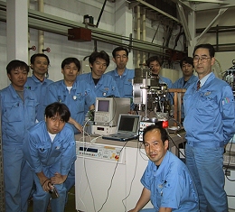
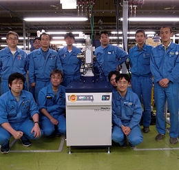
The FR-E, the previous model of the famed FR series, was replaced by the FR-X (photo at right) in 2012. Over 100 units of the FR-E
series (including FR-E+) shipped to customers during its production. Recently,
FR-E Serial No. 002, originally delivered in 2002, came back to the Haijima
factory. The customer wanted the latest technology and highest flux in their
home laboratory so purchased the FR-X and returned Serial No. 002, which was
still running perfectly. Thanks for a great job of more than 10 years! FR-E
Serial No. 002 (as shown at the left in a picture from 2002) will be exhibited in Rigaku's Yamanashi factory as part of its corporate history museum.
By the way, FR-E Serial No. 001 is still working hard at the customer site. As with all Rigaku X-ray sources, longevity and reliability are part of the instrument's DNA.
Chen Yang
Tokyo, Japan
Ask for more information.
Lab spotlight:
Stephen H. White
Stephen H. White, Ph.D.
Professor, Physiology & Biophysics
School of Medicine, University of California, Irvine
Dept. of Physiology and Biophysics
Irvine, CA 92697
Located in the Department of Physiology and Biophysics in the School of Medicine of the University of California at Irvine, White's research group works on biophysical problems related to the folding and stability of membrane proteins. They are part of the UCI Structural Biology and Molecular Biophysics graduate program. Their research is supported by the National Institute of General Medical Sciences and the National Institute of Neurological Disorders and Stroke.
Their research interests include membrane protein folding and stability, energetics of protein-bilayer interactions, experimentally determined hydrophobicity scales, translocon-assisted folding of membrane proteins, structure of fluid lipid bilayers, and MD simulations of lipid bilayers and membrane proteins.
Useful link: Membrane Protein Explorer
Membrane Protein Explorer (MPEx)
is a tool for exploring the topology and other features of membrane proteins based upon thermodynamic and biological principles. Developed in the Stephen H. White lab at UC Irvine, it is designed for examination of membrane protein sequences using hydropathy-plot methods popularized by Kyte and Doolittle (1982). That is, it is based on sliding-window analysis that represents an amino acid sequence as a sequence of numbers representing physical or statistical properties of the amino acid sequence. The most common physical property is hydropathy, which is based upon the free energy of partitioning of amino acids between water and membrane.
This version of MPEx uses two types of hydropathy scales: Experiment-based whole-residue partitioning scales determined in this laboratory and experiment-based biological partitioning scales determined in collaboration with the von Heijne laboratory at Stockholm University. The whole-residue partitioning scales predict with considerable accuracy the transmembrane (TM) segments of membrane proteins of known structure as shown by Jayasinghe et al. (2001). The biological scale utilizes current knowledge of the code the Sec61 translocon to identify TM segments. The algorithm is the same as the one used by von Heijne laboratory on their ΔGpred server, but here it is implemented as a Java Web Start application.
Selected
recent crystallographic papers
Destabilization of the Homotetrameric Assembly of 3-Deoxy-d-Arabino-Heptulosonate-7-Phosphate Synthase from the Hyperthermophile Pyrococcus furiosus Enhances Enzymatic Activity. Nazmi, Ali Reza; Schofield, Linley R.; Dobson, Renwick C.J.; Jameson, Geoffrey B.; Parker, Emily J. Journal of Molecular Biology. Feb2014, Vol. 426 Issue 3, p656-673. 18p. http://dx.doi.org/10.1016/j.jmb.2013.11.008.
Diffuse X-Ray Scattering to Model Protein Motions. Wall, Michael E.; Adams, Paul D.; Fraser, James S.; Sauter, Nicholas K. Structure. Feb2014, Vol. 22 Issue 2, p182-184. 3p. http://dx.doi.org/10.1016/j.str.2014.01.002.
A Structure of a Collagen VI VWA Domain Displays N and C Termini at Opposite Sides of the Protein. Becker, Ann-Kathrin A.; Mikolajek, Halina; Paulsson, Mats; Wagener, Raimund; Werner, Jorn M. Structure. Feb2014, Vol. 22 Issue 2, p199-208. 10p. http://dx.doi.org/10.1016/j.str.2013.06.028.
Electron Density Sharpening as a General Technique in Crystallographic Studies. Liu, Chang; Xiong, Yong. Journal of Molecular Biology. Feb2014, Vol. 426 Issue 4, p980-993. 14p. http://dx.doi.org/10.1016/j.jmb.2013.11.014.
Structure of sugar-bound LacY. Kumar, Hemant; Kasho, Vladimir; Smirnova, Irina; Finer-Moore, Janet S.; Kaback, H. Ronald; Stroud, Robert M. Proceedings of the National Academy of Sciences of the United States of America. 2/4/2014, Vol. 111 Issue 5, p1784-1788. 5p. http://dx.doi.org/10.1073/pnas.1324141111.
Structure-function relationships of membrane-associated GT-B glycosyltransferases. Albesa-Jove, David; Giganti, David; Jackson, Mary; Alzari, Pedro M; Guerin, Marcelo E. Glycobiology. Feb2014, Vol. 24 Issue 2, p108-124. 17p. http://dx.doi.org/10.1093/glycob/cwt101.
Crystal structures of IspF from Plasmodium falciparum and Burkholderia cenocepacia: comparisons inform antimicrobial drug target assessment. O'Rourke, Patrick E. F.; Kalinowska-Tuscik, Justyna; Fyfe, Paul K.; Dawson, Alice; Hunter, William N. BMC Structural Biology. 2014, Vol. 14 Issue 1, p2-27. 26p. http://dx.doi.org/10.1186/1472-6807-14-1.
The 1.59 Å resolution structure of the minor pseudopilin EpsH of Vibrio cholerae reveals a long flexible loop. Raghunathan, Kannan; Vago, Frank S.; Grindem, David; Ball, Terry; Wedemeyer, William J.; Bagdasarian, Michael; Arvidson, Dennis N. BBA - Proteins & Proteomics. Feb2014, Vol. 1844 Issue 2, p406-415. 10p. http://dx.doi.org/10.1016/j.bbapap.2013.11.013.
X-ray Crystallographic and Fluorometric Analysis of the Interactions of Rhein to Human Serum Albumin. Li, Mei; Lee, Philbert; Zhang, Yao; Ma, ZhiYuan; Yang, Feng; Zhou, ZuPing; Wu, XiaoYang; Liang, Hong. Chemical Biology & Drug Design. Feb2014, Vol. 83 Issue 2, p167-173. 7p. http://dx.doi.org/10.1111/cbdd.12208.
Purification, crystallization and X-ray crystallographic studies of flagellin from Pseudomonas aeruginosa. Song, Wan Seok; Hong, Minsun; Yoon, Sung-il. Acta Crystallographica: Section F, Structural Biology Communications. Feb2014, Vol. 70 Issue 2, p200-202. 3p. http://dx.doi.org/10.1107/S2053230X13034286.
Approaches to automated protein crystal harvesting. Deller, Marc C.; Rupp, Bernhard. Acta Crystallographica: Section F, Structural Biology Communications. Feb2014, Vol. 70 Issue 2, p133-155. 23p. http://dx.doi.org/10.1107/S2053230X14000387.
Structural biology: Zooming in on nuclear logistics. Krasteva, Petya V. Nature Methods. Feb2014, Vol. 11 Issue 2, p126-127. 2p. 1 Illustration. http://dx.doi.org/10.1038/nmeth.2827.
Inhibition of master transcription factors in pluripotent cells induces early stage differentiation. Debojyoti De; Myong-Ho Jeong; Young-Eun Leem; Svergun, Dmitri I.; Wemmer, David E.; Jong-Sun Kang; Kyeong Kyu Kim; Sung-Hou Kim. Proceedings of the National Academy of Sciences of the United States of America. 2/4/2014, Vol. 111 Issue 5, p1778-1783. 6p. http://dx.doi.org/10.1073/pnas.1323386111.
Solubility of recombinant Src homology 2 domains expressed in E. coli can be predicted by TANGO. Andersen, Thorny Cecilie; Lindsjo, Kjersti; Dahl Hem, Cecilie; Koll, Lise; Kristiansen, Per Eugen; Skjeldal, Lars; Andreotti, Amy H.; Spurkland, Anne. BMC Biotechnology. 2014, Vol. 14 Issue 1, p1-21. 21p. http://dx.doi.org/10.1186/1472-6750-14-3.
Crystal structures of the human Dysferlin inner DysF domain. Sula, Altin; Cole, Ambrose R.; Yeats, Corin; Orengo, Christine; Keep, Nicholas H. BMC Structural Biology. 2014, Vol. 14 Issue 1, p2-21. 20p. http://dx.doi.org/10.1186/1472-6807-14-3.
Structural and clinical implications of amino acid substitutions in α-l-iduronidase: Insight into the basis of mucopolysaccharidosis type I. Saito, Seiji; Ohno, Kazuki; Maita, Nobuo; Sakuraba, Hitoshi. Molecular Genetics & Metabolism. Feb2014, Vol. 111 Issue 2, p107-112. 6p. http://dx.doi.org/10.1016/j.ymgme.2013.10.005.
Getting CAD in Shape: The Atomic Structure of Human Dihydroorotase Domain. Hermoso, Juan A. Structure. Feb2014, Vol. 22 Issue 2, p179-181. 3p. http://dx.doi.org/10.1016/j.str.2014.01.005.
X-ray structure, thermodynamics, elastic properties and MD simulations of cardiolipin/dimyristoylphosphatidylcholine mixed membranes. Boscia, Alexander L.; Treece, Bradley W.; Mohammadyani, Dariush; Klein-Seetharaman, Judith; Braun, Anthony R.; Wassenaar, Tsjerk A.; Klosgen, Beate; Tristram-Nagle, Stephanie. Chemistry & Physics of Lipids. Feb2014, Vol. 178, p1-10. 10p. http://dx.doi.org/10.1016/j.chemphyslip.2013.12.010.
Mapping transitions between healthy and pathological lesions in human breast tissues by diffraction enhanced imaging computed tomography (DEI-CT) and small angle x-ray scattering (SAXS). Conceicao, A.L.C.; Antoniassi, M.; Geraldelli, W.; Poletti, M.E. Radiation Physics & Chemistry. Feb2014, Vol. 95, p313-316. 4p. http://dx.doi.org/10.1016/j.radphyschem.2013.02.025.
X-Ray Structure of Acid-Sensing Ion Channel 1-Snake Toxin Complex Reveals Open State of a Na+ Selective Channel. Baconguis, Isabelle; Bohlen, Christopher J.; Goehring, April; Julius, David; Gouaux, Eric. Cell. Feb2014, Vol. 156 Issue 4, p717-729. 13p. http://dx.doi.org/10.1016/j.cell.2014.01.011.
Book
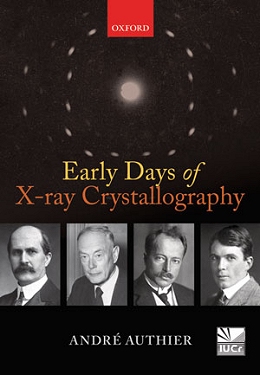
review: Early Days of X-ray Crystallography
by André Authier
Oxford University Press, Oxford, 2013, 441 pages, ISBN 978-0-19-965984-5.
We crystallographers have just finished celebrating the 100th anniversary of the first diffraction experiment, which took place in 1912, and are now celebrating the International Year of Crystallography. Thus, this book could not have been published at a more opportune moment. It is clear that Authier has labored long and hard on this book and, as a result, has done a good job presenting a history of crystallography. He set out to describe the early days of X-ray crystallography and succeeds nicely.
Intriguingly, Authier chose not to follow a strict timeline, instead starting off with a short description of the Laue and Bragg experiments of 1912 and the significance of the actual wavelength of X-rays and the scale of crystal lattices. To pique the reader's interest, Authier provides a list of all of the crystallography related Nobel prizes, thus demonstrating the importance of the method to science.
The next two chapters provide background on the concepts of a space lattice and the dual nature of light, because both are important in understanding the diffraction experiment at its core. Authier then takes readers through the history of the discovery of X-rays by Röntgen in 1895.
All this sets the stage for a detailed history of the original diffraction experiment by von Laue in 1912. In 1913, W. L. Bragg started using the diffraction experiment to determine the structure of simple materials, such as diamond and sodium chloride, which laid the groundwork for our current understanding of the solid state. Authier then explains the X-ray experiment as a branch of optics in the context of the period from 1913 to about 1931, covering kinematic and dynamical theory in easy-to-understand language.
In the final part of the book, Authier highlights the work done by the luminaries of structural studies up to the early fifties, including Pauling, Hull, Bragg, Astbury, along with many other chemists, metallurgists, material scientists and biologists.
In Chapter 11, Authier travels back to ancient Greece, describing the history of crystallography up to 1783. Chapter 12 starts in 1784 with the space lattice concepts of Haüy, who solved the structure of calcite by logic, and ends with the derivation of the 230 space groups by Fedorv, Schoenflies and Barlow in the 1890s. Along the way we learn about the contributions of many others, including but not limited to Miller and Bravais. Given all this, a more appropriate title for the book might have been Early Days of Crystallography.
Some of the concepts are explained with mathematics, but the equations could be skipped by the lay reader and there would be no loss of the history presented. The book is well referenced, with detailed footnotes and short inset biographies of people discussed in the text. It was actually quite nice to learn more about some of the less famous contributors to our science. There is an extensive set of references and a full index.
Unfortunately, there are a few typos in this edition. There is a misspelling of Dorothy Hodgkin's maiden name, Crowfoot, and a wrongly assigned Nobel Prize. One equation has an error in placement of a parenthesis, but it does not change the result. I will leave it to others to catch any other errors and report them to the author.
Joseph D. Ferrara, Ph.D.
Chief Science Officer
|