Background
X-ray diffraction measurements of enamel and dentin, which are the major tissues of a tooth, indicate that they are mostly made of hydroxyapatite. It is not very well known that external treatments, such as laser irradiation, may alter the structure of these components. X-ray microdiffraction is useful to observe the localized influence of laser irradiation on the enamel.
Investigation
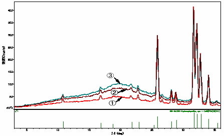
Microdiffraction measurements with ø100 μm X-ray beam were performed on three positions of a section of enamel (labeled 1, 2, 3 in the picture below) using a D/MAX RAPID II diffractometer. The measurement conditions were: X-ray generator output: 40 kV, 36 mA; target: Cu; exposure time: 300sec.
The resulting diffraction patterns indicate that hydroxylapatite polycrystals, the major component of the enamel, became amorphous—not only at the irradiated surface but deeper into the tooth. The amorphous component was largest on the surface (3).

laser irradiation at three labeled measurement points.
Center: Two-dimensional X-ray diffraction data.
Right: Tooth schematic (Ø100 µm each at measurement
sites 1, 2, and 3)
Professor Fumio Hirota of The Nippon Dental University supplied the sample and the data
Reference:
F. Hirota et.al., Lasers in Dentistry, Revolution of Dental Treatment in the New Millennium, (Elsevier Science B.V.) pp.301-311 (2003)