What Are In Vivo Applications?
3D Computed tomography for preclinical small animal studies
Rigaku introduces the latest technology in X-ray computed tomography (CT) for laboratory animals. This in vivo 3D CT instrument allows samples to be monitored for long periods of time.
Cone-shaped X-ray beam for high-speed imaging
This CT exposes a cone-shaped X-ray beam onto the 2D detector. The rotation arm rotates 360° around a sample to acquire full 3D data. This provides 17-second high-speed imaging.
High-resolution microfocus X-ray source
90 kV (maximum X-ray output) and 5 micrometer-focused X-ray source provide high-resolution images that allow users to study sections as small as 20 micrometers.
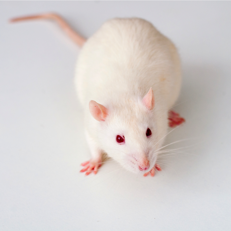
In vivo study
A sample can be periodically monitored in viable condition. This improves experimental efficiency dramatically.

Contact Us
Whether you're interested in getting a quote, want a demo, need technical support, or simply have a question, we're here to help.