Application Note B-XRI1002
Introduction
In food science, observing the internal structure of materials is one of the basic methods to explore the factors determining texture and taste. An electron microscope, such as SEM or TEM, is often used to observe the internal structure of foods. However, pre-processing such as freezing, drying, staining and slicing is necessary to observe the internal structure, so the images are often deformed or contaminated. An X-ray microscope can observe the internal structure without destroying the sample. A Cu source is especially appropriate for visualizing the internal structure with high contrast, even for foods composed of light elements. Here, we illustrate an example of observing the internal structure of string cheese with an X-ray microscope and quantitatively evaluating the thickness of the fibrous part related to the texture of string cheese by 3D image processing.
Measurements and results
A 3D image of a piece of string cheese having a diameter of about 2 mm is obtained from computed tomography (CT) images of 600 sections measured for 2.4 seconds per slice. Figure 1(a)-(c) show orthogonal images sliced from different directions. The center of the intersection on each image represents the same coordinates. White and black colors show high- and low-density regions. The string cheese includes irregularly distributed high-density ingredients of spherical shapes considered to be salt, and low-density ingredients of irregular shape assumed to be oil and water. The fibrous part shown in the side and front view has a net pattern in the top view. The fibrous part was extracted from the CT image of cheese by 3D image processing and was colored by thickness. As a result, the thickness of the fibrous part was 1 to 11 μm in size as shown in Figure 2(b). The 3D image colored by thickness reveals clearly that the fibrous part is aggregated milk proteins lined up in the vertical direction, not shaped like a single fiber (Figure 2(a)). X-ray CT imaging enables the observation of the internal structure of materials at the micrometer scale without pretreatment or destruction of the material. The shape of each component can be visualized and its quantitative result is easily obtained from 3D imaging analysis.
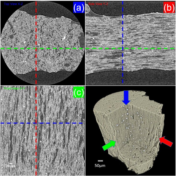
Figure 1. Sliced CT image of string cheese (a) top view (b) side view (c) front view
Figure 2. (a) 3D view of the fibrous part colored by thickness and (b) thickness histogram
